
Molecular structure of the cell membrane. Cell (plasma) membrane, its main functions. History of cell membrane research
Cytoplasm- an obligatory part of the cell, enclosed between the plasma membrane and the nucleus; is divided into hyaloplasm (the main substance of the cytoplasm), organelles (permanent components of the cytoplasm) and inclusions (temporary components of the cytoplasm). Chemical composition cytoplasm: the basis is water (60-90% of the total mass of the cytoplasm), various organic and inorganic compounds. The cytoplasm has an alkaline reaction. Feature cytoplasm of a eukaryotic cell - constant movement ( cyclosis). It is detected primarily by the movement of cell organelles, such as chloroplasts. If the movement of the cytoplasm stops, the cell dies, since only by being in constant motion can it perform its functions.
Hyaloplasma ( cytosol) is a colorless, slimy, thick and transparent colloidal solution. It is in it that all metabolic processes take place, it ensures the interconnection of the nucleus and all organelles. Depending on the predominance of the liquid part or large molecules in the hyaloplasm, two forms of hyaloplasm are distinguished: sol- more liquid hyaloplasm and gel- thicker hyaloplasm. Mutual transitions are possible between them: the gel turns into a sol and vice versa.
Functions of the cytoplasm:
- combining all cell components into a single system,
- environment for the passage of many biochemical and physiological processes,
- environment for the existence and functioning of organelles.
Cell membranes
Cell membranes limit eukaryotic cells. In each cell membrane, at least two layers can be distinguished. The inner layer is adjacent to the cytoplasm and is represented by plasma membrane(synonyms - plasmalemma, cell membrane, cytoplasmic membrane), over which the outer layer is formed. In an animal cell it is thin and is called glycocalyx(formed by glycoproteins, glycolipids, lipoproteins), in a plant cell - thick, called cell wall(formed by cellulose).
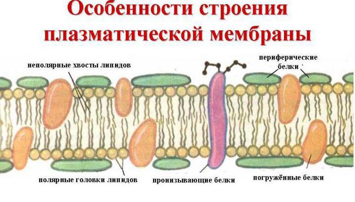
All biological membranes have common structural features and properties. It is currently generally accepted fluid mosaic model of membrane structure. The basis of the membrane is a lipid bilayer formed mainly by phospholipids. Phospholipids are triglycerides in which one fatty acid residue is replaced by a phosphoric acid residue; the section of the molecule containing the phosphoric acid residue is called the hydrophilic head, the sections containing the fatty acid residues are called the hydrophobic tails. In the membrane, phospholipids are arranged in a strictly ordered manner: the hydrophobic tails of the molecules face each other, and the hydrophilic heads face outward, towards the water.
In addition to lipids, the membrane contains proteins (on average ≈ 60%). They determine most of the specific functions of the membrane (transport of certain molecules, catalysis of reactions, receiving and converting signals from the environment, etc.). There are: 1) peripheral proteins(located on the outer or inner surface of the lipid bilayer), 2) semi-integral proteins(immersed in the lipid bilayer to varying depths), 3) integral, or transmembrane, proteins(penetrate the membrane through, contacting both the outer and internal environment cells). Integral proteins are in some cases called channel-forming or channel proteins, since they can be considered as hydrophilic channels through which polar molecules pass into the cell (the lipid component of the membrane would not let them through).
A - hydrophilic phospholipid head; B - hydrophobic phospholipid tails; 1 - hydrophobic regions of proteins E and F; 2 — hydrophilic regions of protein F; 3 - branched oligosaccharide chain attached to a lipid in a glycolipid molecule (glycolipids are less common than glycoproteins); 4 - branched oligosaccharide chain attached to a protein in a glycoprotein molecule; 5 - hydrophilic channel (functions as a pore through which ions and some polar molecules can pass).
The membrane may contain carbohydrates (up to 10%). The carbohydrate component of membranes is represented by oligosaccharide or polysaccharide chains associated with protein molecules (glycoproteins) or lipids (glycolipids). Carbohydrates are mainly located on the outer surface of the membrane. Carbohydrates provide receptor functions of the membrane. In animal cells, glycoproteins form a supra-membrane complex, the glycocalyx, which is several tens of nanometers thick. It contains many cell receptors, and with its help cell adhesion occurs.
Molecules of proteins, carbohydrates and lipids are mobile, capable of moving in the plane of the membrane. The thickness of the plasma membrane is approximately 7.5 nm.
Functions of membranes
Membranes perform the following functions:
- separation of cellular contents from the external environment,
- regulation of metabolism between the cell and the environment,
- dividing the cell into compartments (“compartments”),
- place of localization of “enzymatic conveyors”,
- ensuring communication between cells in the tissues of multicellular organisms (adhesion),
- signal recognition.
The most important membrane property— selective permeability, i.e. membranes are highly permeable to some substances or molecules and poorly permeable (or completely impermeable) to others. This property underlies the regulatory function of membranes, ensuring the exchange of substances between the cell and the external environment. The process of substances passing through the cell membrane is called transport of substances. There are: 1) passive transport- the process of passing substances without energy consumption; 2) active transport- the process of passage of substances that occurs with the expenditure of energy.
At passive transport substances move from an area of higher concentration to an area of lower, i.e. along the concentration gradient. In any solution there are solvent and solute molecules. The process of moving solute molecules is called diffusion, and the movement of solvent molecules is called osmosis. If the molecule is charged, then its transport is also affected by the electrical gradient. Therefore, people often talk about an electrochemical gradient, combining both gradients together. The speed of transport depends on the magnitude of the gradient.
The following types of passive transport can be distinguished: 1) simple diffusion- transport of substances directly through the lipid bilayer (oxygen, carbon dioxide); 2) diffusion through membrane channels— transport through channel-forming proteins (Na +, K +, Ca 2+, Cl -); 3) facilitated diffusion- transport of substances using special transport proteins, each of which is responsible for the movement of certain molecules or groups of related molecules (glucose, amino acids, nucleotides); 4) osmosis— transport of water molecules (in all biological systems the solvent is water).
Necessity active transport occurs when it is necessary to ensure the transport of molecules across a membrane against an electrochemical gradient. This transport is carried out by special carrier proteins, the activity of which requires energy expenditure. The energy source is ATP molecules. Active transport includes: 1) Na + /K + pump (sodium-potassium pump), 2) endocytosis, 3) exocytosis.

Operation of Na + /K + pump. For normal functioning, the cell must maintain a certain ratio of K + and Na + ions in the cytoplasm and in the external environment. The concentration of K + inside the cell should be significantly higher than outside it, and Na + - vice versa. It should be noted that Na + and K + can diffuse freely through the membrane pores. The Na + /K + pump counteracts the equalization of the concentrations of these ions and actively pumps Na + out of the cell and K + into the cell. The Na + /K + pump is a transmembrane protein capable of conformational changes, as a result of which it can attach both K + and Na +. The Na + /K + pump cycle can be divided into the following phases: 1) addition of Na + from the inside of the membrane, 2) phosphorylation of the pump protein, 3) release of Na + in the extracellular space, 4) addition of K + from the outside of the membrane , 5) dephosphorylation of the pump protein, 6) release of K + in the intracellular space. Almost a third of all energy required for cell functioning is spent on the operation of the sodium-potassium pump. In one cycle of operation, the pump pumps out 3Na + from the cell and pumps in 2K +.
Endocytosis- the process of absorption of large particles and macromolecules by the cell. There are two types of endocytosis: 1) phagocytosis- capture and absorption of large particles (cells, parts of cells, macromolecules) and 2) pinocytosis— capture and absorption of liquid material (solution, colloidal solution, suspension). The phenomenon of phagocytosis was discovered by I.I. Mechnikov in 1882. During endocytosis, the plasma membrane forms an invagination, its edges merge, and structures delimited from the cytoplasm by a single membrane are laced into the cytoplasm. Many protozoa and some leukocytes are capable of phagocytosis. Pinocytosis is observed in intestinal epithelial cells and in the endothelium of blood capillaries.
Exocytosis- a process reverse to endocytosis: the removal of various substances from the cell. During exocytosis, the vesicle membrane merges with the outer cytoplasmic membrane, the contents of the vesicle are removed outside the cell, and its membrane is included in the outer cytoplasmic membrane. In this way, hormones are removed from the cells of the endocrine glands; in protozoa, undigested food remains are removed.
Go to lectures No. 5"Cell theory. Types of cellular organization"
Go to lectures No. 7“Eukaryotic cell: structure and functions of organelles”
Cell membrane- this is the cell membrane that performs the following functions: separation of the contents of the cell and the external environment, selective transport of substances (exchange with the environment external to the cell), the place where some biochemical reactions, association of cells into tissues and reception.
Cell membranes are divided into plasma (intracellular) and external. The main property of any membrane is semi-permeability, that is, the ability to pass only certain substances. This allows for selective exchange between the cell and the external environment or exchange between cell compartments.
Plasma membranes are lipoprotein structures. Lipids spontaneously form a bilayer ( double layer), and membrane proteins “float” in it. The membranes contain several thousand different proteins: structural, transporters, enzymes, etc. Between the protein molecules there are pores through which hydrophilic substances pass (the lipid bilayer prevents their direct penetration into the cell). Glycosyl groups (monosaccharides and polysaccharides) are attached to some molecules on the surface of the membrane, which are involved in the process of cell recognition during tissue formation.
Membranes vary in thickness, usually ranging from 5 to 10 nm. The thickness is determined by the size of the amphiphilic lipid molecule and is 5.3 nm. A further increase in membrane thickness is due to the size of membrane protein complexes. Depending on external conditions (cholesterol is the regulator), the structure of the bilayer can change so that it becomes more dense or liquid - the speed of movement of substances along the membranes depends on this.
Cell membranes include: plasma membrane, karyolemma, membranes of the endoplasmic reticulum, Golgi apparatus, lysosomes, peroxisomes, mitochondria, inclusions, etc.
Lipids are insoluble in water (hydrophobicity), but soluble in organic solvents and fats (lipophilicity). The composition of lipids in different membranes is not the same. For example, the plasma membrane contains a lot of cholesterol. The most common lipids in the membrane are phospholipids (glycerophosphatides), sphingomyelins (sphingolipids), glycolipids and cholesterol.
Phospholipids, sphingomyelins, glycolipids consist of two functional various parts: hydrophobic nonpolar, which does not carry charges - “tails” consisting of fatty acids, and hydrophilic, containing charged polar “heads” - alcohol groups (for example, glycerol).
The hydrophobic part of the molecule usually consists of two fatty acids. One of the acids is saturated, and the second is unsaturated. This determines the ability of lipids to spontaneously form bilayer (bilipid) membrane structures. Membrane lipids perform the following functions: barrier, transport, protein microenvironment, electrical resistance of the membrane.
Membranes differ from each other in their set of protein molecules. Many membrane proteins consist of regions rich in polar (charge-bearing) amino acids and regions with nonpolar amino acids (glycine, alanine, valine, leucine). Such proteins in the lipid layers of membranes are located so that their non-polar sections are, as it were, immersed in the “fat” part of the membrane, where the hydrophobic sections of lipids are located. The polar (hydrophilic) part of these proteins interacts with the lipid heads and faces the aqueous phase.
Biological membranes have common properties:
membranes are closed systems that do not allow the contents of the cell and its compartments to mix. Violation of the integrity of the membrane can lead to cell death;
superficial (planar, lateral) mobility. In membranes there is a continuous movement of substances across the surface;
membrane asymmetry. The structure of the outer and surface layers is chemically, structurally and functionally heterogeneous.
If you find an error, please highlight a piece of text and click Ctrl+Enter.
The cell membrane is the planar structure from which the cell is built. It is present in all organisms. Her unique properties ensure the vital activity of cells.
Types of membranes
There are three types of cell membranes:
- external;
- nuclear;
- organelle membranes.
The outer cytoplasmic membrane creates the boundaries of the cell. It should not be confused with the cell wall or membrane found in plants, fungi and bacteria.
The difference between a cell wall and cell membrane in a much greater thickness and the predominance of the protective function over the exchange function. The membrane is located under the cell wall.
The nuclear membrane separates the contents of the nucleus from the cytoplasm.
TOP 4 articleswho are reading along with this
Among the cell organelles there are those whose shape is formed by one or two membranes:
- mitochondria;
- plastids;
- vacuoles;
- Golgi complex;
- lysosomes;
- endoplasmic reticulum (ER).
Membrane structure
By modern ideas The structure of the cell membrane is described using a fluid mosaic model. The basis of the membrane is a bilipid layer - two levels of lipid molecules forming a plane. On both sides on bi lipid layer protein molecules are located. Some proteins are immersed in the bilipid layer, some pass through it.

Rice. 1. Cell membrane.
Animal cells have a complex of carbohydrates on the surface of the membrane. When studying a cell under a microscope, it was noted that the membrane is in constant motion and is heterogeneous in structure.
The membrane is a mosaic in both a morphological and functional sense, since its different sections contain different substances and have different physiological properties.
Properties and Functions
Any border structure carries out protective and exchange functions. This applies to all types of membranes.
The implementation of these functions is facilitated by such properties as:
- plastic;
- high ability to recover;
- semi-permeability.
The property of semi-permeability is that some substances are not allowed to pass through the membrane, while others pass freely. This is how the control function of the membrane is carried out.
Also, the outer membrane provides communication between cells due to numerous outgrowths and the release of an adhesive substance that fills the intercellular space.
Transport of substances across the membrane
Substances enter through the outer membrane in the following ways:
- through pores with the help of enzymes;
- through the membrane directly;
- pinocytosis;
- phagocytosis.
The first two methods are used to transport ions and small molecules. Large molecules enter the cell by pinocytosis (in liquid state) and phagocytosis (in solid form).

Rice. 2. Scheme of pino- and phagocytosis.
The membrane wraps around the food particle and locks it into the digestive vacuole.
Water and ions pass into the cell without energy expenditure, through passive transport. Large molecules move by active transport, consuming energy resources.
Intracellular transport
From 30% to 50% of the cell volume is occupied by the endoplasmic reticulum. This is a kind of system of cavities and channels that connects all parts of the cell and ensures orderly intracellular transport of substances.

Rice. 3. EPS drawing.
Thus, a significant mass of cell membranes is concentrated in the ER.
What have we learned?
We found out what a cell membrane is in biology. This is the structure on which all living cells are built. Its significance in the cell is to: delimit the space of organelles, the nucleus and the cell as a whole, ensuring the selective flow of substances into the cell and nucleus. The membrane consists of lipid and protein molecules.
Test on the topic
Evaluation of the report
Average rating: 4.7. Total ratings received: 485.
It's no secret that all living beings on our planet are made up of cells, these countless "" organic matter. The cells, in turn, are surrounded by a special protective shell - a membrane, which plays a very important role in the life of the cell, and the functions of the cell membrane are not limited to just protecting the cell, but represent a complex mechanism involved in the reproduction, nutrition, and regeneration of the cell.
What is a cell membrane
The word “membrane” itself is translated from Latin as “film,” although a membrane is not just a kind of film in which a cell is wrapped, but a combination of two films connected to each other and having different properties. In fact, the cell membrane is a three-layer lipoprotein (fat-protein) membrane that separates each cell from neighboring cells and the environment, and carries out controlled exchange between cells and environment, this is the academic definition of what a cell membrane is.
The importance of the membrane is simply enormous, because it not only separates one cell from another, but also ensures the cell’s interaction with both other cells and the environment.
History of cell membrane research
An important contribution to the study of the cell membrane was made by two German scientists Gorter and Grendel back in 1925. It was then that they managed to conduct a complex biological experiment on red blood cells - erythrocytes, during which scientists obtained so-called “shadows”, empty shells of erythrocytes, which they stacked in one stack and measured the surface area, and also calculated the amount of lipids in them. Based on the amount of lipids obtained, scientists came to the conclusion that they are precisely contained in the double layer of the cell membrane.
In 1935, another pair of cell membrane researchers, this time Americans Daniel and Dawson, after a series of long experiments, established the protein content in the cell membrane. There was no other way to explain why the membrane had such a high surface tension. Scientists have cleverly presented a model of a cell membrane in the form of a sandwich, in which the role of bread is played by homogeneous lipid-protein layers, and between them, instead of oil, there is emptiness.
In 1950, with the advent electronic theory Daniel and Dawson were able to confirm this with practical observations - in micrographs of the cell membrane, layers of lipid and protein heads and also the empty space between them were clearly visible.
In 1960, the American biologist J. Robertson developed a theory about the three-layer structure of cell membranes, which for a long time was considered the only correct one, but with further development science, doubts began to arise about its infallibility. So, for example, from the point of view, it would be difficult and labor-intensive for cells to transport the necessary nutrients through the entire “sandwich”
And only in 1972, American biologists S. Singer and G. Nicholson were able to explain the inconsistencies in Robertson’s theory using a new fluid-mosaic model of the cell membrane. In particular, they found that the cell membrane is not homogeneous in its composition, moreover, it is asymmetrical and filled with liquid. In addition, cells are in constant motion. And the notorious proteins that are part of the cell membrane have different buildings and functions.

Properties and functions of the cell membrane
Now let's look at what functions the cell membrane performs:
The barrier function of the cell membrane is the membrane as a real border guard, standing guard over the boundaries of the cell, delaying and not allowing harmful or simply inappropriate molecules to pass through.
Transport function of the cell membrane - the membrane is not only a border guard at the cell gate, but also a kind of customs checkpoint; useful substances are constantly exchanged with other cells and the environment through it.
Matrix function - it is the cell membrane that determines the location relative to each other and regulates the interaction between them.
Mechanical function - is responsible for limiting one cell from another and, at the same time, for correctly connecting cells to each other, for forming them into a homogeneous tissue.
The protective function of the cell membrane is the basis for building the cell's protective shield. In nature, an example of this function can be hard wood, a dense peel, a protective shell, all due to the protective function of the membrane.
Enzymatic function is another important function performed by certain proteins in the cell. For example, thanks to this function, the synthesis of digestive enzymes occurs in the intestinal epithelium.
Also, in addition to all this, cellular exchange occurs through the cell membrane, which can take place in three different reactions:
- Phagocytosis is a cellular exchange in which membrane-embedded phagocyte cells capture and digest various nutrients.
- Pinocytosis is the process of capture by the cell membrane of liquid molecules in contact with it. To do this, special tendrils are formed on the surface of the membrane, which seem to surround a drop of liquid, forming a bubble, which is subsequently “swallowed” by the membrane.
- Exocytosis is a reverse process when a cell releases a secretory functional fluid to the surface through the membrane.

Structure of the cell membrane
There are three classes of lipids in the cell membrane:
- phospholipids (which are a combination of fats and),
- glycolipids (a combination of fats and carbohydrates),
- cholesterol
Phospholipids and glycolipids, in turn, consist of a hydrophilic head, into which two long hydrophobic tails extend. Cholesterol occupies the space between these tails, preventing them from bending; all this, in some cases, makes the membrane of certain cells very rigid. In addition to all this, cholesterol molecules organize the structure of the cell membrane.
But be that as it may, the most important part of the structure of the cell membrane is protein, or rather different proteins that play different important roles. Despite the diversity of proteins contained in the membrane, there is something that unites them - annular lipids are located around all membrane proteins. Annular lipids are special structured fats that serve as a kind of protective shell for proteins, without which they simply would not work.
The structure of the cell membrane has three layers: the basis of the cell membrane is a homogeneous liquid bilipid layer. Proteins cover it on both sides like a mosaic. It is proteins, in addition to the functions described above, that also play the role of peculiar channels through which substances that are unable to penetrate through the liquid layer of the membrane pass through the membrane. These include, for example, potassium and sodium ions; for their penetration through the membrane, nature provides special ion channels in cell membranes. In other words, proteins ensure the permeability of cell membranes.
If we look at the cell membrane through a microscope, we will see a layer of lipids formed by small spherical molecules on which proteins swim as if on the sea. Now you know what substances make up the cell membrane.
Cell membrane video
And finally, an educational video about the cell membrane.

When writing the article, I tried to make it as interesting, useful and high-quality as possible. I will be grateful for any feedback And constructive criticism in the form of comments to the article. You can also write your wish/question/suggestion to my email. [email protected] or on Facebook, sincerely the author.
This article is available at English language – .
The branch of biology called cytology studies the structure of organisms, as well as plants, animals and humans. Scientists have found that the contents of the cell, which is located inside it, are built quite complex. It is surrounded by the so-called surface apparatus, which includes the outer cell membrane, supra-membrane structures: the glycocalyx and also microfilaments, pelicule and microtubules that form its submembrane complex.
In this article we will study the structure and functions of the outer cell membrane included in the surface apparatus various types cells.
What functions does the outer cell membrane perform?
As described earlier, the outer membrane is part of the surface apparatus of each cell, which successfully separates its internal contents and protects cellular organelles from unfavorable conditions external environment. Another function is to ensure metabolism between cellular contents and tissue fluid, so the outer cell membrane transports molecules and ions entering the cytoplasm, and also helps remove waste and excess toxic substances from the cell.

Structure of the cell membrane
The membranes, or plasma membranes, of different types of cells differ greatly from each other. Mainly, chemical structure, as well as the relative content of lipids, glycoproteins, proteins in them and, accordingly, the nature of the receptors located in them. The external one, which is determined primarily by the individual composition of glycoproteins, takes part in the recognition of environmental stimuli and in the reactions of the cell itself to their actions. Some types of viruses can interact with proteins and glycolipids of cell membranes, as a result of which they penetrate the cell. Herpes and influenza viruses can be used to build their protective shell.
And viruses and bacteria, the so-called bacteriophages, attach to the cell membrane and dissolve it at the point of contact using a special enzyme. Then a viral DNA molecule passes into the resulting hole.
Features of the structure of the plasma membrane of eukaryotes
Let us recall that the outer cell membrane performs the function of transport, that is, the transfer of substances in and out of it into the external environment. To carry out such a process, a special structure is required. Indeed, the plasmalemma is a permanent, universal system of surface apparatus. This is a thin (2-10 Nm), but quite dense multilayer film that covers the entire cell. Its structure was studied in 1972 by scientists such as D. Singer and G. Nicholson, and they also created a fluid-mosaic model of the cell membrane.
The main chemical compounds that form it are ordered molecules of proteins and certain phospholipids, which are embedded in a liquid lipid medium and resemble a mosaic. Thus, the cell membrane consists of two layers of lipids, the non-polar hydrophobic “tails” of which are located inside the membrane, and the polar hydrophilic heads are facing the cell cytoplasm and the intercellular fluid.
The lipid layer is penetrated by large protein molecules that form hydrophilic pores. It is through them that they are transported aqueous solutions glucose and mineral salts. Some protein molecules are found on both the outer and inner surfaces of the plasmalemma. Thus, on the outer cell membrane in the cells of all organisms that have nuclei, there are carbohydrate molecules bound covalent bonds with glycolipids and glycoproteins. The carbohydrate content in cell membranes ranges from 2 to 10%.

The structure of the plasmalemma of prokaryotic organisms
The outer cell membrane in prokaryotes performs similar functions to the plasma membranes of cells of nuclear organisms, namely: perception and transmission of information coming from the external environment, transport of ions and solutions into and out of the cell, protection of the cytoplasm from foreign reagents from the outside. It can form mesosomes - structures that arise when the plasma membrane is invaginated into the cell. They may contain enzymes involved in metabolic reactions of prokaryotes, for example, DNA replication and protein synthesis.
Mesosomes also contain redox enzymes, and photosynthetics contain bacteriochlorophyll (in bacteria) and phycobilin (in cyanobacteria).
The role of outer membranes in intercellular contacts
Continuing to answer the question of what functions the outer cell membrane performs, let us dwell on its role in. In plant cells, pores are formed in the walls of the outer cell membrane, which pass into the cellulose layer. Through them, the cytoplasm of the cell can exit to the outside; such thin channels are called plasmodesmata.

Thanks to them, the connection between neighboring plant cells is very strong. In human and animal cells, the contact points between adjacent cell membranes are called desmosomes. They are characteristic of endothelial and epithelial cells, and are also found in cardiomyocytes.
Auxiliary formations of the plasmalemma
Understanding how plant cells differ from animal cells is helped by studying the structural features of their plasma membranes, which depend on the functions of the outer cell membrane. Above it in animal cells there is a layer of glycocalyx. It is formed by polysaccharide molecules associated with proteins and lipids of the outer cell membrane. Thanks to the glycocalyx, adhesion (sticking together) occurs between cells, leading to the formation of tissues, therefore it takes part in the signaling function of the plasmalemma - recognizing environmental stimuli.
How is the passive transport of certain substances carried out across cell membranes?
As mentioned earlier, the outer cell membrane is involved in the process of transporting substances between the cell and the external environment. There are two types of transport through the plasmalemma: passive (diffusion) and active transport. The first includes diffusion, facilitated diffusion and osmosis. The movement of substances along a concentration gradient depends, first of all, on the mass and size of molecules passing through the cell membrane. For example, small nonpolar molecules easily dissolve in the middle lipid layer of the plasmalemma, move through it and end up in the cytoplasm.

Large molecules of organic substances penetrate into the cytoplasm with the help of special carrier proteins. They have species specificity and, when connecting with a particle or ion, passively transfer them across the membrane along a concentration gradient without energy expenditure (passive transport). This process underlies such a property of the plasmalemma as selective permeability. During the process, the energy of ATP molecules is not used, and the cell saves it for other metabolic reactions.
Active transport of chemical compounds through the plasmalemma
Since the outer cell membrane ensures the transfer of molecules and ions from the external environment into the cell and back, it becomes possible to remove dissimilation products, which are toxins, outside, that is, into the intercellular fluid. occurs against a concentration gradient and requires the use of energy in the form of ATP molecules. It also involves carrier proteins called ATPases, which are also enzymes.
An example of such transport is the sodium-potassium pump (sodium ions move from the cytoplasm into the external environment, and potassium ions are pumped into the cytoplasm). Epithelial cells of the intestines and kidneys are capable of it. Varieties of this transfer method are the processes of pinocytosis and phagocytosis. Thus, having studied what functions the outer cell membrane performs, it can be established that heterotrophic protists, as well as cells of higher animal organisms, for example, leukocytes, are capable of the processes of pino- and phagocytosis.
Bioelectric processes in cell membranes
It has been established that there is a potential difference between the outer surface of the plasma membrane (it is positively charged) and the wall layer of the cytoplasm, which is negatively charged. It was called the resting potential, and it is inherent in all living cells. And nervous tissue not only has a resting potential, but is also capable of conducting weak biocurrents, which is called the process of excitation. The outer membranes of nerve cells-neurons, receiving irritation from receptors, begin to change charges: sodium ions massively enter the cell and the surface of the plasmalemma becomes electronegative. And the near-wall layer of the cytoplasm, due to an excess of cations, receives a positive charge. This explains why the outer cell membrane of the neuron is recharged, which causes the conduction of nerve impulses that underlie the excitation process.